


Muscular system
Muscles can either contract or relaxExpanded state
Muscles connect to bones using tendons and ligaments connect bone to bone
Types of muscles:
There are 3 types of muscles.
Cardiac muscles:
Muscles present in your heart
Smooth muscles:
Carries out most involuntary processes
Most of it is present in the digestive system, urinary system and blood vessels but some are also present in other parts of the body
Skeletal muscles:
They are also called striated muscles
They carry out voluntary actions
640 skeletal muscles are present in humans
Anatomy of skeletal muscles:
Courtesy:National Cancer Institute
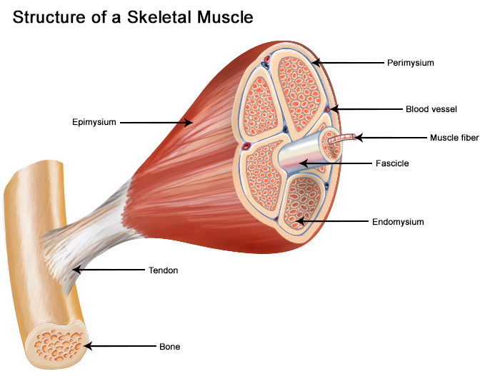
The thick middle portion of the muscle is called the muscle belly
This tapers off into tendons on either end
Tendons are fibrous proteins, mostly collagen
The muscles stretch from one joint to another, allowing bones to move relative to other bones
Connective tissue outside the muscle called fascia separate muscles from each other
Each muscles is surrounded by a sheath made of connective tissue called the epimysium
Inside the muscle there are compartments containing bundles of muscle fibers, these bundles are called fascicles
Each fascicle is surrounded by connective tissue called perimysium
Each muscle fiberMuscle cell is wrapped in a sheath called endomysium
Epimysium,perimysium,endomysium collectively form the tendon or a thin sheet of connective tissue called the aponeurosis
The aponeurosis also helps in connecting muscles to bone
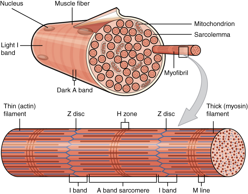
Each muscle cell contains multiple nuclei and are formed by a bunch of cellsProgenitor cells fusing together
Muscle cells contain organelles called myofibril
Myofibrils are composed of thick, thin and elastic filamentsCollectively called myofilaments repeating in segments
Each segment is called a sarcomere
Terms related to muscle fibers:
T-tubules:
Part of the cell membrane that runs perpendicular to the direction of the myofibril.
This allows the action potential to reach the SR.
The ends of the SR are called the terminal cisterna
The region where around the t-tubule is called the triad region as it contains a terminal cisterna on either side of the t-tubule
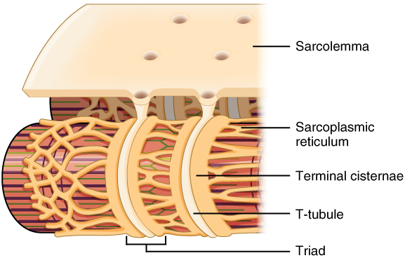
Sarcolemma:
The cell membrane of a muscle fiber
Z-disc/Z-line:
2 dark colored bands present on either sides of the sarcomere.
I-band:
The region of the sarcomere where only the thin filament is present.
This region is lighter than the surrounding region
A-band:
The region of the sarcomere where the thin and thick filaments overlap.
This region is darker than the I-band
H-zone:
With in the A-band there is a lighter section where only the thick filament is present.
It is as wide as the gap between the thin filaments.
M-line:
The H-zone is bisected by a dark line called the M-line.
Working of skeletal muscles:
The thick myofilament is mainly made of a protein called myosin
The thin myofilament is mainly made of a protein called actinElastic myofilaments are mainly made of the protein titin
The actin is surrounded by tropomyosin and troponin
Tropomyosin are made of 2 strands which wrap around actin giving it structure and prevents myosin from grabbing itPrevents muscle contraction until a proper signal
Each tropomyosin has a troponin attached to it in the grooves between actin filaments
There is a special version of the endoplasmic reticulum(ER) called the sarcoplasmic reticulum(SR)
The SR is around each sarcomere and has calcium pumps attached, pumping in calcium creating a concentration gradient
Sliding filament model:
Each myosin is already energised with the hydrolysis of an ATP
Each sarcomere has a motor neuron near it that can trigger it
The neuron triggers an action potential in the muscle fiber
This action potential travels along the membrane of the fiber and then flow into special folds called t-tubules
When the signal reaches the SR, the SR releases calcium ions into the myofibrils
The calcium ions bind with troponin, causing it to move itself and the tropomyosin away from the binding sites on the actin and allows the myosin to grab the actin
When the myosin grabs the actin then the energy from the ATP hydrolysis releases causing the myosin to move the actin towards the center of the sarcomereTowards other actin
For the myosin and actin to be put back into their original positions, they need an ATP to attach the head to the myosin and release energy and turn into ADPThis means that the natural state of a muscle is contracted
Then the SR pumps calcium back inside it and the troponin and tropomyosin go back to their original positions